Review on Sheep and Goat Pox
Received Date: September 09, 2021 Accepted Date: October 09, 2021 Published Date: October 11, 2021
doi: 10.17303/ejmrc.2021.3.102
Citation:Yonas Dawle (2021) Review on Sheep and Goat Pox. Eur J Med Res Clin Trials 1: 1-11.
Abstract
Sheep and Goat Pox is a highly contagious viral disease that causes detrimental effect on sheep and goat in many parts of the world. Sheep and Goat Pox viruses are in the Pox viridae family and Capripox virus genus is the causative agent. The virus can survive for several weak in oral and nasal secretion after infection and also can live in scabs that have fallen off the animal for several months. Spread can also occur from contact with contaminated materials and through skin abrasions produced iatrogenically or by insects. The diseases are most commonly transmitted through direct contact, indirect contact and mechanically by the vectors. Up on entry of virus into the host it replicates in local tissue and spread to different parts of the body as viremia causing pox lesions on skin (hair less area), lung, liver and kidney. The systemic signs include fever, conjunctivitis, rhinitis, lymphadenopathy, anorexia and depression. The skin lesions follow a typical pox virus development from a macule to papule and appear as small red patches usually around the mouth, on the head, under the tail and between the legs. The mucous membranes of the eyes, nose, mouth, vulva and prepuce may be necrotic ulcerated and all the body lymph nodes are swollen and enlarged. The disease has major impact on the economy with average morbidity and mortality rates of 50 and 100%, respectively. The morbidity and mortality of the disease is more severe in lambs, kids and exotic breed. Heavy economic losses in sheep pox outbreaks are due to the high mortality, damage of skin, abortions and loss of market value of the affected animals. Diagnosis of the diseases is depends on clinical signs, laboratory confirmation and post mortem examinations. Use of antibiotics for control of secondary bacterial complications, vaccination, limitation of animal movement and their products are important ways to control the disease. Enhanced awareness on appropriate bio-security measures to be undertaken by livestock keepers can greatly reduce the impact of this disease.
Keywords:Sheep and Goat Pox Virus; Goat, Sheep; Sheep and Goat Pox
Introduction
Capri poxviruses are the most important poxviruses of animals, causing diseases in sheep, goats or cattle. It is responsible for some of the most economically significant diseases of domestic ruminants in Africa and Asia [1].Various strains of Capri pox virus are responsible for the disease and these are antigenically and serologically indistinguishable from strains causing sheep pox and goat pox but distinct at the genetic level [2].The genus Capri pox virus, member of the pox viridae consists of lumpy skin disease virus, goat pox virus and sheep pox virus [3].
CaPV genomes are very similar to each other, averaging no less than 96% nucleotide identity over their entire length. SPPV, GTPV, and LSDV contain the same repertoire of orthologous genes, with the exception that SPPV and GTPV lack nine LSDV genes with likely CaPV virulence and host range functions. SPPV and GTPV genomes sequenced are phylogenetically distinct from each other and from LSDV, and they contain species-specific nucleotide differences that may be associated with aspects of host range. Relatively few genomic changes in SPPV and GTPV vaccine viruses account for viral attenuation [4].
In general, Capripox viruses (CaPV) are considered to be very host-specific (Babiuk, et al., 2009) [5]. In addition to the isolate Kenya Sheep and Goat Pox (KSGP O-240), only a few other SPPV and GTPV strains have been known to affect both sheep and goats (Yan, et al., 2012) [6]. It causes highly infectious disease in sheep and goats where the disease is less commonly seen in indigenous breeds in area where it’s endemic as compared with exotic breeds. It is transmitted by direct contact, indirect contact with infected object or fomites and through insect that can mechanically transmit the diseases. Up on establishment of infection in the host it can causes highly devastating systemic viremia which is characterized by widespread skin eruption, fever, generalized papules or nodules, vesicles (rarely) on hairless area of the skin, internal lesions in the lungs, respiratory and gastrointestinal mucosa and cause death of the animals (Abd-Elfatah, et al., 2018) [7]. The diseases is distributed in most part of the world where it is commonly seen in Middle East, Africa (north of the equator), the Indian subcontinent, much of central Asia, and in South-Eastern Europe where sporadic outbreaks occur. Recently outbreaks have been recorded in Kazakhstan, Mongolia, Azerbaijan, Turkey, Greece and Bulgaria (Beard, et al., 2010; Gelaye, et al., 2013; Tuppurainen, et al., 2017) [8,9,47]. In Africa the number of countries affected by sheep and goat pox virus were showing an increase in the trend of the diseases particularly for three consecutive years before 2011. However, the number of countries affected by sheep and goat pox virus remarkably decreased after 2011 in which twelve countries were reported from twenty six countries affected by the diseases in 2010 that indicate the reduction of the diseases by 46% which is happen due to national intervention. In Africa among the endemic countries that recorded high number of outbreak in 2011 Ethiopia were the first where high outbreak is recorded (AU IBAR, 2011) [10]. Sheep and goat pox are among the most important diseases of sheep and goats in Ethiopia following Peste des petits ruminants (PPR) and Contagious caprine pleuropneumonia (CCPP) that affect small ruminants entailing a huge economic loss and listed as trans-boundary disease of animal affecting the economy of the country (Befikadu and Endale, 2017) [11]. A review on past and current aspects indicated that the disease distributed in all regions of Ethiopia and economically important due to production loss and direct death (Yune and Abdela, 2017) [12].
Therefore, the objective of this seminar paper is to review the status, epidemiology and economic importance of sheep and goat pox.
Etiology
Sheep and Goat Pox viruses are in the Pox viridae family, Chordopoxvirinae subfamily and Capripox virus genus (Buller, et al., 2005) [3]. Capripox viruses are large brick-shaped, double stranded DNA viruses, morphologically indistinguishable from Orthopox viruses, measuring 295 by 265 nm (Kitching and Smale, 1986) [13]. The virion is covered in short tubular elements which give it a different appearance from Orf virus, which is more oval in shape and covered in a continuous filament. Poxviruses have a very stable genome which is demonstrated by DNA restriction patterns of isolates collected in 1959 appearing the same as 1986 isolates, indicating very little change in the genome over time (Kitching, et al., 1989) [14]. However, sheep-and goat pox viruses will have subtle genetic changes to their genomes over time which can be identified using genome sequencing. In addition, there is evidence for recombination events occurring between strains of capripox virus in the field, as has also been seen in vitro, and this could result in changes in host range or virulence (Gershon, et al., 1989) [15].
In natural conditions, sheep and goat poxviruses are pathogenic exclusively for the ovine and caprine species respectively. In addition to the isolate Kenya Sheep and Goat Pox virus (KSGPV O-240, only a few other Sheep Pox Virus (SPPV) and Goat Pox Virus (GTPV)) strains have been known to affect both sheep and goats (Yan, et al., 2012). The major difference between the African and the Middle Eastern and Indian SPP and GTP strains seems to be the wider host range of the African isolates. The Kenya sheep-1 (KS-1) strain is derived from the attenuated KSGP O-240 vaccine strain (Davies, 1976) [16].
As with other poxviruses, sheep- and goat pox is susceptible to sunlight and detergents containing lipid solvents, but in dark environmental conditions, such as contaminated animal sheds, it can persist for many months. The source of environmental contamination occurs from infected animals shedding the virus (Essbauer, et al., 2007) [17].
Epidemiology
Distribution
Sheep and Goat Pox are prevalent in parts of, central Asia, Africa except in South Africa, and the Middle Eastern countries. Goat pox is first reported in 879 in Norway and was later observed in Macedonia during the First World War. Capripox virus is found in the Middle East, in Africa north of equator India, Pakistan, Turkey and Iran. Recent outbreak was occurred in 2008 and 2009 in Mogolia. Another outbreak have occurred in 2008 and 2009 is in Greece and Kazakhstan and Azerbaijan respectively. In Vietnam goat pox has been introduced in 2005. The outbreak of goat pox was occurred in Chinese Taipei in 2008 and in 2010 the disease reoccurred and was declared endemic (OIE, 2012) [18]. In Ethiopia, the disease is distributed in all regions (Webbs, 1980) [19].
The occurrence of SPP and GTP as reported to OIE in 2010, 2013 and 2015 respectively, Countries reporting SPP and GTP are highlighted in red colour as revealed on the figures, the disease is propagating in Africa, Middle East and Asia.
Transmission
The virus of sheep and got pox is highly contagious. Virus enters via respiratory tract and transmission is mostly by aerosol through contact with infected animal or fomite. Vectors like, Stomoxys, Calcitrans and tsetse fly can transmit virus mechanically (Webbs, 1980). Most experimental transmission and pathogenesis studies have used intradermal inoculation of the virus. Disseminated infection of the skin following either experimental intradermal inoculation or after respiratory infection is the result of viraemia and subsequent systemic viral spread to the skin (Afshar, et al., 1986) [21]. The virus can survive for several weak in oral and nasal secretion after infection and also can live in scabs that have fallen off the animal for several months. Spread can also occur from contact with contaminated materials and through skin abrasions produced iatrogenically or by insects (AHA, 2011; Radostits, et al., 2006) [22,23].
Risk Factor
Pathogen risk factor: The poxviruses are thought to have prolonged survival in environment and inactivated by drying, freezing, thawing, and remain viable for months in the lyophilized state. But its sensitive to 1% of formalin and extreme PH. can remain infectious for up to six months in sheep pens, and may also be found on the wool or hair for as long as three months after infection (Sharma, et al.,1988) [24]. Capripox virus is highly stable in normal environment condition and can survive for prolonged time, with or without susceptible animal. They are inactivated by sun light and heat, but can survive in cool dark environment for up to 6 month (Davies, 1981) [25].
Host risk factor: Group of Sheep and goat of all age, breed and sex are susceptible to sheep and goat pox. In areas where sheep pox is enzootic, imported breeds such as Merinos or some European breeds may show greater susceptibility than the native stock. Sheep and goat pox infect only sheep and goat and have no zoonosis. Wild ungulate is not reservoir for this disease (ESGPIP, 2009) [26]. Capripox virus can affect sheep, goat and cattle. Virus of goat pox is highly host-specific, infecting only goats, but from isolate-to-isolate host specificity varies. It is possible that the host preference shown by different strains is due to their adaptation to the presence of either sheep or goat alone in a limited geographical area. Isolates of Capripox virus are not host-specific; cattle, goats, and sheep who have recovered from infection with Capripox virus isolates from a heterologous host hav immune to any challenge with a virulent homologous virus (Kitching, et al., 1989) [14]. There are two types of sheep pox virus (Singari, 1990) [27], in which, one affects both sheep and goats (Kenyan sheep and goat (KSG) strain while the other is host specific. Recent records indicate that strains of sheep pox do pass between sheep and goats, although most cause more severe disease in sheep. Recombination also occurs between strains of SPV producing a spectrum, showing intermediate host preferences and a range of virulence (OIE, 2000) [28].
Environment risk factor: Environmental determinants play a great role in the occurrence of sheep and goat pox. It had impact on the agent, host and vectors as well as interaction between them. These predisposing factors have a great role in maintenance of Stomoxys, Calcitrans and the tsetse fly to susceptible animals which are the vectors for transmission of disease (Webbs, 1980) [19].
Prevalence
The outbreak of Capripox virus was first reported in 1936 in India, and since then, frequent outbreaks have been reported throughout the country in almost all the states where sheep and goats are reared. The occurrence of the disease is usually observed throughout the year, however, most frequently seen during the rainy season (Garner, et al., 2000) [29]. Mortality in young animals can exceed 50%. A recent report revealed that the morbidity and mortality rates in the flock were 18.4% and 6.3%, respectively (Hemadri & Hiremath, 2011) [30]. Exotic and young animals are highly susceptible (Bhanuprakash, et al., 2006) [1].
An outbreak of sheep pox occurred in April 2017 on a local breed of sheep in Kafr Shalshamoun, Menya Al Qamh, Sharkia, Egypt include 85 sheep (56 adults one and 29 young lambs aged 3 to 6 months old). The total percentage of diseased animals was 23.5% and the total percentage of dead animals was 8.2%. The incidence was higher than recorded by Mondal, et al. (2004) [31] reported that the incidence of infection was 18.4% during outbreak of SPPV occurred in December 2001 in Jammu, India and also higher than outbreak of SPPV that occurred in Greece 2007 with incidence of 8.45% (Mangana, et al., 2008) [32] and other detected in Sudan (18.9%) and in India (7.2%) by Nour, et al. (2012) [33] and Selvaraju (2014) [34], respectively. But less than other reported during an outbreak of SPPV among sheep occurred in Ningxia Hui, China among sheep during November 2011, the incidence of the disease was 29.2% (Zhu, et al., 2013) [35]. In this investigation the mortality rate was 8.2% which higher than the percentage recorded by Mondal, et al. (2004) [31] in which the percentage of death was 6.3% and less than recorded by Ammar, et al. (1999) [36] detected that large number of sheep at El-Karada, Sakha and El-Gemiza in Kafr El- Sheikh Province showed skin eruptions on wool less areas of the skin with mortality rate 68.4%, diagnosis referring to SPPV infection, Zhu, et al. (2013) [35] 14.6%, and by El- Sabagh, et al. (2014) [37] during outbreak of sheep pox disease occurred in Al-Hassa province of Saudi Arabia during the period between the winter and the spring 2013, mortality was15%. This variation in both incidence rate and mortality rate may be attributed to difference in the study time, total number of examined animals, study localities and method of rearing.
In Ethiopia, the prevalence of skin defect due to sheep and goat pox disease identified respectively in sheep and goat was 44% &56% from Modjo export tannery (Berhanu, et al., 2011) [38], 19% & 17% from Bahar dar tannery (Azene, et al., 2015) [39], 1.2% &15.5% from Sheba Tigray tannery (Kahsay, et al., 2015) [40] and 4.2% &5.3% from Mojo and Addis Ababa tanneries (Bisrat, 2014) [41]. During outbreak investigation in central Ethiopia (Mulo, Sululta, Adea-berga, Yaya-gulale and Tiyo) from the total of 603 local sheep and 109 goats examined, 216 sheep were showed pox lesions where as 31 goats were positive for pox lesion. From the affected groups, 60 sheep and 4 goats were died. Overall, 35.82%, 9.95% and 27.7% morbidity, mortality and case fatality where observed in sheep respectively. Whereas 28.44%, 3.66% and 12.9% morbidity, mortality and case fatality rate respectively, were observed in goats (Aberaham, 2018) [42].
Prevalence
The pathogenesis depends on both the virus isolate and the host. In addition, virus replication is apparently correlated with the severity of clinical disease, i.e. the highest levels of virus replication occur in association with severe disease (Babiuk, et al., 2009) [5]. Capripox viruses have a predilection for epithelial cells of the skin and lungs (Bowden, et al., 2008) [43]. Incubation period of sheep pox is 4-8 days and of that of goat pox is 4-15 days. After it enters, goat pox virus replicates locally in the tissues. Since the virus is epitheliotropic, it will infest the epithelium tissues of the organism. Based on a trial conducted, on the 7th day post-inoculation, the virus titer reached to its peak. The virus spread to the regional lymph nodes, after 3-4 days of primary viremia. The viremia spread in the body and affected spleen, lungs and liver. The virus inhaled may also cause lungs lesions. In skin nodules from 7 to 14 days after inoculation, the virus titters persisted and decreased with the development of serum antibodies. Within 24 hours of the appearance of generalized papules, affected animals develop conjunctivitis, rhinitis and enlargement of all the superficial lymph nodes, in particular the prescapular lymph nodes. Excessive salivation can also occur after infection (Kitching, et al., 1989) [14].
There are five stages in the development of poxvirus infection. Roseola stage is stage in which Skin lesions typically begin with small red spots within three days of infection which is followed by papules. The affected animals are febrile at this stage. The second stage of pox lesion is Papules which develops after 3 days of roseola stage. Nodular skin lesions that are developed from roseola stage (red spots) those are hard during palpation. Papules within 5-6 days are changed to vesicles and known as vesicular stage. Pustular stage develops after 3 days of vesicular stage. As stated by researchers, the last stage of pox lesion is scab. Study has shown that, quantitative analysis using real-time PCR and isolation of the pathogenesis of Sheep pox virus and Goat pox virus in their respective hosts’ revealed high viral loads in skin (Hurisa, et al., 2018) [20].
Clinical Signs and Necropsy Findings
The disease is more severe in lambs and kids than in adult animals. Some very young lambs and kids may die before exhibiting signs of the disease (Gitao, et al., 2017) [44]. The clinical signs of sheep pox and goat pox can be either malignant or benign. The malignant form of sheep pox and goat pox is mostly common in lamb. Affected lambs may die without observable pox lesion. Fevers which peak at 40-42 °C, dyspnoea and occulonasal discharge and pox lesion on unwooled skin are manifested in malignant form of sheep pox and goat pox. The diseases are more severe in young animals than adults. In benign form of sheep pox and goat pox only skin lesions occur particularly under the tail. This form of sheep pox and goat pox is common in adults. There is no systemic reaction and the animal recovers in 3-4 weeks. Abortion and secondary pneumonia are complications. Lesions may be seen on the vulva, perineum, nostril and mucous membranes of the mouth. If lesion is present in the lung acute respiratory distress occurs (Ausvetplan, 1996) [45].
Most affected animals become weak with no appetite. Skin lesions appear as small red patches usually around the mouth, on the head, under the tail and between the legs. The centres of the patches become depressed and turn greyish in colour due to necrosis. These patches form blisters that break becoming open sores that soon develop scabs. The ruptured lesions often form a hard black scab. These skin lesions render infected sheep prone to fly strike and the scabs may persist for several weeks (Gitao, et al., 2017) [44]. It considered that the clinical signs of severe sheep pox are pathognomic. Animals often have laboured breathing due to blisters inside the respiratory tract and lungs. Lesions in the mouth, nose and eyes can cause discharge become necrotic and ulcerate. Nodules in the intestines can cause diarrhoea (Kitching, 2007) [46].
At post mortem, high percentage of affected animals may develop pneumonia, which grossly appears as multiple foci of sub-pleural consolidation and oedema. In addition, pox lesions can sometimes be found on many serosal surfaces. The mucous membranes of the eyes, nose, mouth, vulva and prepuce may be necrotic ulcerated and all the body lymph nodes are swollen and enlarged. Lymph nodes draining infected areas are enlarged up to eight times normal size, swollen with body fluids and may be congested and haemorrhagic. Kitching (2007) commented that the large number of hard, pale, sometimes haemorrhagic lesion found throughout the lobes of the lung are usually the most obvious and the most likely cause of death. Some authors have however; found the lesions to be soft rather than hard (Watt and Scrivener, 2014). At necropsy, affected sheep would show widely dispersed discrete, generally circular, slightly raised, firm 1-2 cm papules on the lung surface, occasionally in the mucosa of the rumen and abomasums and rarely, small (2-5 mm) lesions in the renal cortex (Watt and Scrivener, 2014) and liver (Kitching (2007) [46, 47].
Economic Importance
The mortality rate of SPP and GTP can sometimes be considerably high particularly amongst exotic breed of sheep and goats, lambs and kids (Tuppurainen, et al., 2017). Where it is the highly contagious SPPV and GTPV are able to cause mortality up to 50% in susceptible flock. Young animals show more severe disease and mortality in lambs and kids may be as high as 100% (Hamouda, et al., 2017) [51].
The disease has major impact on the economy with average morbidity and mortality rates of 50 and 100%, respectively. The effect is such that it would take 6 years for a flock or herd to recover from an outbreak with average income losses up to 30–43% of total annual revenue depending on flock type and owners actions. The level of impact varies from country-to-country both qualitatively and quantitatively. SGPV is one of the animal bioterrorist agents as it causes high morbidity and mortality, has potential for rapid spread, potential to cause serious socio-economic or public health consequences and is of major importance in the international trade of animals and animal products. Sheep pox virus is one of the 15 animal pathogens listed by Animal World Health Organization (OIE) and 23 by Animal and Plant Health Inspection Agency which can be used as an animal biological warfare agent (USDA, 2002) [56].
It is suggested that goat pox is the most important of all pox diseases of domestic animals causing high mortality in kids and significant economic losses. In one study performed in Israel, it was found that SPP and GTP, in endemic areas, are associated with significant production losses because of reduced milk yield (up to 30 %), high mortality in lambs (95 %), decreased average conception rate (32 %) in the year following the outbreak, decreased weight gain, increased abortion rates (3 %), damage to wool and hides and increased susceptibility to pneumonia and fly strike (Yeruham, et al., 2007) [57]. In India, where the disease is endemic, a survey conducted in Maharashtra state revealed that the disease has a major impact on the economy, with average morbidity and mortality rates of 63.5 and 49.5 %, respectively. The effect is such that it would take six years for a flock or herd to recover from an outbreak, with average income losses up to 30–43 % of the total annual revenue depending on flock type and owners’ actions. State wide, around 5000 flocks and herds are affected annually, incurring losses in the range of Indian Rubies 107.5 million (Fakri, et al., 2015) [50].
In a study conducted in Ethiopia to evaluate the impact of major cattle, sheep and goat diseases impacting on the Hides and skins industry, whose exports average a yearly value of $52,160,000 USD, it was found that SGP had a prevalence of 5.94%, impacted accordingly to losses incurred by the country in that sector of the economy (Teshome and Derso, 2015) [54]. In Ethiopia, generally the annual economic loss due to disease, mortality, reduced reproductive and productive performance of small ruminants was estimated by 150 million USD where there is 5-7 million sheep and goats die each year due to disease where among the most common disease Sheep and goat pox is the one leads to loss (Tsegaye, et al., 2013) [55].
Diagnosis
Sheep and goat Pox can be diagnosed based on observable clinical sign like, fever, dyspnea and pox lesion in different parts of the unwoolen skin. Clinical pathology and species of affected host are also important in the diagnosis of this disease. Epidemiology of the disease is also important in diagnoses of sheep and goat pox. As the virus of sheep and goat pox are very closely related it’s indistinguishable by serologically (Yune and Abdela, 2017). Sheep or goat pox should be suspected in animals with the characteristic full-thickness skin lesions, fever, and lymphadenitis. Laboratory procedures for the diagnosis of sheep and goat pox include observation of the virus by electron microscopy (morphology is characteristic) and virus isolation (identification is by immune fluorescence or immune peroxidise occur in the AGID test) with Parapoxvirus. Serology is also useful; antibodies can be found one week after the skin lesions appear. Serologic tests include virus neutralization, AGID, indirect immune fluorescence, ELISA, and immune blotting (Western blotting). Virus neutralization is the most specific serological test, but is not sensitive enough to detect infections in all animals. Cross-reactions with other viruses are seen in the AGID and indirect immune fluorescence tests. Histopathologic lesions are also characteristic (Gitao, et al., 2017).
Sheep and goat pox may have similar clinical sign with Contagious ecthyma (Orf), bluetongue, Parasitic pneumonia, Caseous lymphadenitis, Insect bites, Dermatophilosis, Sheep scab, Mange and Photsensitization, Peste des petits ruminants (Yune and Abdela, 2017).
Treatment, Prevention and Control
Sheep and goat pox has no effective treatment therefore in order to overcome the loss due to the disease treatment approach is dependent on control of secondary bacterial infection which can be achieved through parenteral administration of broad spectrum antibiotic to avoid complication of bacterial infections. For early recovery of the animal cleaning of infected areas with weak solution of potassium permanganate (1:10000) and topically application of antibiotic ointment is important for skin lesion, keeping the animal in well ventilated enclosure, provision of balanced diet is helpful and in case if animals are unable to feed 10% glucose saline should be given parentally (Senthilkumar and Thirunavukkarasu, 2010; CABI, 2015) [53].
Active mass vaccination to SGP may induce strong herd immunity that can effectively control the disease. Single vaccination is considered as enough for providing lifelong strong immunity (Bhanuprakash, 2011) [49]. Among the live attenuated vaccine, SGPV vaccine and subunit formulations have been used experimentally in enzootic and outbreak areas as vaccines against Sheep pox, Goat pox, and Lumpy Skin Disease (Chibssa, et al., 2019). However, vaccine-induced disease and vaccine failure are some of the challenges that need to improve CaPV vaccines through understanding of the genetic base of virus virulence and host range to produce high efficacy vaccine (Tulman, et al., 2002; Chibssa, et al., 2018) [48].
Control of the disease, once it has entered, is usually by early detection and notification, prompt movement restriction of animals, culling affected and in-contact animals, and ring vaccination , early detection and notification, prompt movement restriction of animals, culling affected and in-contact animals, and ring vaccination with a vaccine (Kitching, 1986) [52]. Routine control measures include the cleaning and disinfection of depopulated premises and establishment of protection and surveillance zones, with a radius of 3 and 10 km, respectively, around the outbreak (Magana, et al., 2008). As noted earlier, uncontrolled movement of infected animals in SGP-endemic areas poses serious difficulties in efficient control of the disease. Therefore, it is essential to vaccinate sheep flocks regularly, on an annual basis, with a safe and efficient vaccine, for the control of this serious and economically important disease in endemic regions (Yeruham, et al., 2007).
Conclusion and Recommendations
Sheep and goat pox is contagious viral diseases which affect sheep and goats causing lesions on the hairless parts of the body and internal organs. The disease distributed most countries of the world and lead to extensive loss in different countries. In Ethiopia, the diseases outbreak occurred each year at different parts of the country. Thus it result in directly or indirectly influence the economy of the country causing detrimental loss in the production and productivity, restriction of live animal export, products and by products and even leads to death of the animal. Control and eradication of sheep and goat pox is challenging due to illegal movement of animas that allow the extension of the disease to different parts of the world, genetic similarity of the virus among species and genus which influence the diagnosis of the disease, lack of early diagnostic test to control outbreaks, the ability of the virus to undergo recombination, vaccine efficacy and failure problems which leads to the reoccurrence of diseases after vaccination. Therefore, based on the above conclusive remarks the following recommendations are forwarded:-
- Awareness should be created on the husbandry and herd management to minimize the transmission of the disease between sheep and goats.
- Thorough control, prevention and eradication program has to be planed and implemented.
- Early diagnostic approach has to be developed to reduce the loss due to the disease outbreak.
- Further studies should be conducted on isolation of Circulating sheep and goat Virus strain and their vaccine matching.
- Bhanuprakash V, Indrani BK, Hosamani M, Singh RK (2006) The current status of sheep pox disease. Comparative Immunology, Microbiology& Infectious Diseases, 29: 27–60.
- Babiuk S, Bowden TR, Boyle DB, Wallace, Kitching RP (2008) Capripoxviruses: an emerging worldwide threat to sheep, goats and cattle. Transboundary and Emerging Diseases, 55: 263-72.
- Center for Agriculture and Biosciences International (CABI) (2015) Sheep and Buller, R., Arif B. and Black D. (2005): Pox viridae in virus taxonomy eight report of the international committee on the taxonomy of viruses, Elsevior Acadamic press, Oxford: 117.
- Tulman ER, Afonso CL, Lu Z, Zsak L, Sur JH,, et al. (2002) The Genomes of Sheep pox and Goat pox Viruses. Journal of Virology, 76: 6054-61.
- Babiuk S, Bowden TR, Parkyn G, Dalman B, Hoa DM,, et al. (2009): Yemen and Vietnam capripox viruses demonstrate a distinct host preference for goats compared with sheep. J Gen Virol 90: 105-14.
- Yan XM, Chu YF, Wu GH, Zhao ZX, Li J,, et al. (2012) An outbreak of sheep pox associated with goat poxvirus in Gansu province of China. Vet Microbiol 156: 425-8.
- Abd-Elfatah EB, El-Mekkawi MF, Bastawecy IM, Fawzi EM (2018) Identification and phylogentic analysis of sheep pox during an outbreak of sheep in Sharkia Governorate, Egypt. Genetics and Molecular Res 17: 1-12.
- Beard PM, Sugar S, Bazarragchaa E, Gerelmaa U, Tserendorj S,, et al. (2010) A description of two outbreaks of Capripox virus disease in Mongolia. Veterinary Microbiology 142: 427-31.
- Gelaye E, Mach L, Kolodziejek J, Grabherr R, Loitsch A,, et al. (2017) A novel HRM assay for the simultaneous detection and differentiation of eight poxviruses of medical and veterinary importance. J Sci Rep 7: 42892.
- African Union Inter African Bureau for Animal Resources (AU-IBAR) (2011) Pan African Animal Health Year book.
- Befikadu, S, Endale T (2017) Major Transboundary Disease of Ruminants and their Economic Effect in Ethiopia. Global Journal of Medical Res 17(2): 27-36.
- Yune N, Abdela N (2017) Epidemiology and Economic Importance of Sheep and Goat Pox: A Review on Past and Current Aspects. J Vet Science Technol 8: 1-5.
- Kitching RP, Smale C (1986) Comparison of the external dimention of capripoxvirus isolates Research in Veterinary Science, 41: 425-7.
- Kitching RP, Bhat PP, Black DN (1989) The characterization of African strains of capripoxviruses. Epidemiology and Infection 102: 335-43.
- Gershon PD, Kitching RP, Hammond JM, Black DN (1989) Poxvirus genetic recombination during natural virus transmission. J General Virology, 70: 485–9.
- Davies FG (1976) Characteristics of a virus causing a pox disease in sheep and goat in Kenya, with observations on the epidemiology and control. J Hyg Camb 76: 163-71.
- Essbauer S, Meyer H, Porsch-Ozcürümez M, Pfeffer M (2007) Long-lasting stability of vaccinia virus (orthopoxvirus) in food and environmental samples. Zoonoses Public Health 54:118-24.
- Manual OIE (2000) Manual of standards for diagnostic tests and vaccines, 4th edn.
- Webbs G (1980) Sheep and goat pox, transmission of capripox viruses by various flies indicated the need for a reassessment of the methods of controlling this disease. Annual Report, Institute for Animal Health, Pirbright, UK.
- Hurisa TT, Jing Z, Jia H, Chen G, He XB (2018) Review on Sheeppox and Goatpox: Insight of Epidemiology, Diagnosis, Treatment and Control Measures in Ethiopia. J Infect Dis Epidemiol 4: 057.
- Afshar A, Bundza A, Myers DJ, Dulac GC, Thomas FC (1986) Sheep pox Experimental studies with a west african isolate. Canadian Veterinary Journal, 27: 301-6.
- Animal Health Australia (AHA) (2011) Disease Strategy Sheep pox and goat pox.
- Radostits OM, Gay CC, Hinchcliff KW (2006) Veterinary Medicine, 10th edn. Saunders: 1430-1.
- Sharma B, Negi BS, Pandey AB, Bandyopadhyay SK, Shankar H (1988) Detection of goat pox antigen and antibody by the CIE test. Tropical Animal Health and Production, 20: 109-13.
- Davies FG (1981) Sheep and Goat pox in Virus diseases of food Animals. London: Academic Press: 733-48.
- Ethiopian Sheep and Goats Productivity Improvement Program (ESGPIP) (2009) Common defects of sheep and goats skin in Ethiopia and their causes. Technical Bulletin 19.
- Singari NA, Moorthy AS, Rama Rao P (1990) Sheep pox. Livest Adviser 15: 40-2.
- OIE Terrestrial Manual (2012) Chapter 2.7.14. Sheep pox and goat pox.
- Garner MG, Sawarkar SD, Brett EK, Edwards JR, Kulkarni VB,, et al. (2000) The extent and impact of sheep pox and goat pox in the state of Maharashtra, India. Tropical Animal Health Production 32: 205–23.
- Hemadri D, Hiremath J (2011) Vision 2030 Project Directorate on Animal Disease Monitoring and Surveillance: 1-30.
- Mondal B, Hosamani M, Dutta TK, Senthilkumar VS (2004) An outbreak of sheep pox on a sheep breeding farm in Jammu, India. Rev Sci tech Off Int Epiz 23: 943-9.
- Mangana O, Kottaridi C, Nomikou K (2008) The epidemiology of sheep pox in Greece from 1987 to 2007. Rev Scient Tech 27: 899-905.
- Nour TAM, Fadol MA, Ahmed AM, Yagoub IA (2012) Studies on the humoral immune response of sheep to Sheep Pox Thermostable Vaccine. The Sudan. J Vet Res 26: 7-12.
- Selvaraju G (2014) Epidemiological measures of disease frequency against Sheep pox. Inter J of scienti Res 3: 2277-8179.
- Zhu XL, Yang F, Li HX, Dou YX (2013) Identification and phylogenetic analysis of a Sheeppox virus isolated from the Ningxia Hui Autonomous Region of China. Genetics and Molecular Res 12: 1670-8.
- Ammar KM, Al-Gaabary MH, Abou-Rawash AA, Foad FM (1999) Clinical, epidemiological and histopathological studies on sheep pox in some farms in Egypt. 5th Sci. Cong., Egyptian Society for Cattle Disease: 60-2.
- El-Sabagh I, Al-Shabebi A, Abu-Elzein E, Zaghawa A (2014) Molecular detection and phylogenetic analysis of Sheeppox virus in Al – Hassa of Eastern Province of Saudi Arabia Advances in Animal and Vet Sci 2: 31-4.
- Berhanu W, Negussie H, Alemu S, Mazengia H (2011) Assessment on major factors that cause skin rejection at Modjo export tannery, Ethiopia. Tropical Animal Health and Production, 43: 989–93.
- Azene D, Fentahun T, Admassu B (2015) Study on the Major Defects That Causes Sheep and Goat Skins Rejection in Bahir Dar Tanning Industry, Ethiopia. Academic Journal of Animal Diseases, 4: 170-6.
- Kahsay T, Negash G, Hagos Y, Hadush B (2015) Pre-slaughter, slaughter and postslaughter defects of skins and hides at the Sheba Tannery and Leather Industry, Tigray region, northern Ethiopia. Onderstepoort J Vet Res 82: 1-7.
- Bisrat GU (2014) Investigation of Major Factors That Cause Skin and Hide Rejection in Ethiopia. Case of Tanneries in Addis Ababa and Modjo Towns. J Africa Leather and Leather Products Advances 1: 35-43.
- Aberaham DA (2018) Outbreak investigation, isolation and molecular Characterization of sheep and goat pox virus in centeral Ethiopia. MSc Thesis of Addis Ababa University, Faculty of Veterinary Medicine, Bishoftu, Ethiopia.
- Bowden TR, Babiuk SL, Parkyn GR, Copps JS, Boyle DB (2008) Capripoxvirus tissue tropism and shedding. Quantitative Study in Experimentally Infected Sheep and Goats. Virology 371: 380-93
- Gitao CG, Mbindyo C, Omani R, Chemweno V (2017) Review of Sheep Pox Disease in Sheep. Department of Veterinary Pathology and Microbiology, University of Nairobi, Kenya.
- AUSVETPLAN (1996) Australian Veterinary Emergency Plan. Disease Strategy Lumpy skin disease.
- Watt B, Scrivener C (2014) Sheep and Goat Pox. Skirting the Issues, The Official Newsletter of the Australian Sheep Veterinarians. Autumn 6-9.
- Tuppurainen ES, Venter EH, Shisler JL, Gari G, Mekonnen GA,, et al. (2017) Review on Capripox virus Diseases: Current Status and Opportunities for Control. Transboundary and Emerging Diseases, 64: 729–45.
- Chibssa TR, Grabherr R, Loitsch A, Settypalli TB, Tuppurainen E,, et al. (2018) A gel-based PCR method to differentiate sheep pox virus field isolates from vaccine strains. Virology J 15: 59.
- Bhanuprakash V (2011) Prospects of control and eradication of capripox from the Indian subcontinent. A perspective Antiviral Res 91: 225-32.
- Fakri F, Ghzal F, Daouam S, Elarkam A, Douieb L,, et al. (2015) Development and field application of a new combined vaccine against PPR and Sheep Pox. Trials in Vaccinology, 4: 33–7.
- Hamouda M, Al-Hizab F, El-Sabagh I (2017) Clinical, pathological and molecular diagnosis of sheep pox virus in Saudi Arabia. Journal of Animal and Plant Sciences 27: 9197.
- Kitching RP (1986) The Control of Sheep and goat pox. Rev Sci Tech off Epiz 5: 503-11.
- Senthil kumar V, Thirunavukkarasu M (2010) Economic losses due to sheep pox in sheep farms in Tamil Nadu. Tamil Nadu Journal of Veterinary and Animal Sciences, 6: 88-94.
- Teshome D, Derso S (2015) Prevalence of Major Skin Diseases in Ruminants and its Associated Risk Factors at University of Gondar Veterinary Clinic, North West Ethiopia. J Veterinar Sci Technol S: S13002.
- Tsegaye D, Belay B, Haile A (2013) Prevalence of Major Goat Diseases and Mortality of Goat in Daro-Labu District of West Hararghe, Eastern Ethiopia. Journal of Scientific and Innovative Res 2: 665-72.
- USDA (2002) Agricultural bioterrorism act of. Fedl Regist; 67: 52383–9.
- Yeruham I, Yadin H, Van Ham M, Bumbarov V, Soham A,, et al. (2007): Economic and epidemiological aspects of an outbreak of sheep pox in a dairy sheep flock. Veterinary Record, 160: 236-7. 1.
FIGURE 1
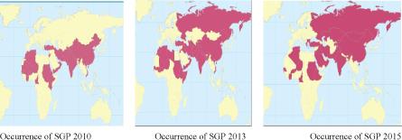
Source: Tuppurainen, et al. (2017); Hursia, et al. (2018) [20].
Figure 1: Global occurrence of SPP and GTP as reported to OIE
FIGURE 2

Figure 2: Lesions in different organs
Lesions in lung (A and B), in kidney (C), wall of abomasums (D), in omentum and wall of rumen and in rumen wall (F).
Figures at a glance