Early Postoperative Topographic changes after laser Asymmetric Keratectomy for the Management of Adverse Effects after Photorefractive Keratectomy
Received Date: September 22, 2020 Accepted Date: October 22, 2020Published Date: October 24, 2020
doi: 10.17303/jooa.2020.4.102
Citation:Ji Sang Min (2020) Early Postoperative Topographic changes after laser Asymmetric Keratectomy for the Management of Adverse Effects after Photorefractive Keratectomy. J Ophthalmol Open Access 1: 1-7.
Abstract
Laser asymmetric keratectomy (LAK) is a biomechanical customization method to avoid and treat conventional laser refractive surgery adverse effects. A 57-year-old male patient had undergone photorefractive keratectomy (PRK) on the left eye to correct -3.50 diopters 27 years ago and had astigmatic change (-1.75 85°) and blurred vision in the left eye from 6 months after PRK. Uncorrected and best-corrected visual acuities were 20/40. On the Orbscan map, the cone of the left cornea was located in the nasal side, the corneal thickness imbalance was severe due to 207µm of the sum of 4 direction differences in corneal thickness. So, he had distorted, asymmetric cornea with irregular astigmatism after PRK. LAK was performed in two stages of ablation. First, after ascertaining the deviations in central symmetry based on the Vision-Up program, the deviations were eliminated. The corneal curvature, which shows myopic shifts due to the corneal ablation procedure, can be predicted by the Vision-Up program. Thus, additional ablation for myopia correction and ablation for refractive errors were performed simultaneously to ensure that the first procedure did not cause a myopic shift. Pre and postoperative two months topographic changes were compared on the Orbscan map especially with the sums of 4 direction corneal thickness deviation and distance between the optic axis and the apex of the posterior cornea. Sum of deviations in corneal thickness (µm) in four directions based on Orbscan maps was markedly decreased from 207µm preoperatively to 138µm postoperatively. The distance between the optic axis and the apex of the posterior cornea was significantly decreased after LAK. The Thinnest point (cone) was located closely in the center of the cornea. Uncorrected visual acuity also markedly increased postoperatively. There were no complaints in blurring. It is clinically important to assess the early topographic changes of the post- LAK technique for the management of adverse effects.
Keywords: Adverse effects; Laser Asymmetric keratectomy (LAK); Deviations in corneal thickness (µm) in four directions based on Orbscan maps
Introduction
Myopic regression or decrease in visual acuity and ocular discomfort have been reported following laser corneal refractive surgery [1-5].
Laser surgeries were attempted to relieve the adverse effects of LASIK or LASEK, but these were not effective [6-9]. Conventional corneal refractive correction methods that focus only on correcting refractive abnormalities are unable to prevent corneal deformation because of the interaction between the intraocular pressure and corneal thickness in patients with a large deviation in corneal thickness, showing asymmetric corneas. Therefore, it has been suggested that only new biomechanical customization methods could prevent postoperative corneal deformation [10-16]. The corneal shape is determined by a complex interaction between its biomechanical properties, such as corneal thickness and stiffness and intraocular pressure [10,11,17,18]. In this regard, recent research [19-21] has focused on postoperative adverse effects in patients with a large sum of deviation in corneal thickness in 4 directions≥80um preoperatively. In the patients with large deviation in corneal thickness (≥80um), increased corneal irregularities and corneal asymmetry, higher blurring scores, lower uncorrected distance visual acuity, and lower efficiency index was shown postoperatively [21], but in cases of a decreasing sum of a total deviation in corneal thickness with asymmetric corneal ablation(from ≥80um to < 80um), adverse effects were markedly decreased postoperatively [21]. Thus, corneal thickness deviation is considered an important factor of corneal asymmetry and a cause of visual abnormalities such as blurring of vision, visual aberrations, and dry eye [19-21].
Recent studies have suggested laser asymmetric keratectomy (LAK) [19, 21] as a method for biomechanical customization. It helps achieve central corneal symmetry with a central point, and such improved corneal morphology is maintained long-term without deterioration of the cornea [19-21].
Therefore, corneal topography is very important to evaluate the early surgical result. The authors performed LAK to relieve adverse effects such as the distorted cornea or blurring after PRK and attempted to report changes in corneal topography two months after LAK.
Case Presentations
Clinical findings and Timeline
A 57-year-old male patient had undergone PRK to correct -3.50 diopters 27 years ago in an eye clinic and had astigmatic changed and blurred vision in the left eye after 6 months. He complained with halos and starburst. There were no underlying diseases such as diabetes. Before LAK, uncorrected visual acuity was 20/40 in the left eye, and the best-corrected visual acuity 20/40 in the left eye.
Diagnostic Assessment
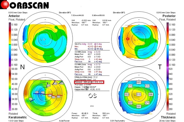
The refraction of the left eye was measured -1.75 85° using an auto refracto keratometer and the intraocular pressure measured with a non-contact tonometer was 14mmHg.The other ocular findings including retina were fine. For pre and postoperative corneal thickness deviation [19, 21], the thickness (µm) was measured at 8 points corresponding to the direction angles (0º, 45º, 90º, 135º, 180º, 225º, 270º, and 315º) at a distance of 2.5 mm from the center of the cornea. The differences in thickness between symmetrically opposed points (0–180º, 45–225º, 90–270º, and 135–315º) were calculated, and the sum of these differences was used in the analysis (Figure 1 and 2). The sum of 4 direction differences in corneal thickness was 207µm in the left eye (Figure 1). The distance [19, 21] between the maximum posterior elevation (best-fit-sphere) and the visual axis was checked by conversion to the distance between the X and Y coordinates of the thinnest point and the center of the cornea on an Orbscan map (Figure 1 and 2). The central cone of the left eye cornea was asymmetrical cornea from the nasal side. The corneal thickness imbalance was severe in the left eye (Figure 1). The corneal irregularities (diopters) in the 3.0-mm and 5.0-mm zone on the Orbscan maps were ±3.2 diopters and ±3.7dioptersin the preoperative LAK. The central corneal thickness was 515um, the pupil diameter was 3.1mm, and the Kappa angle was 1.97°.
Therapeutic Intervention
Patients with refractive error and corneal thickness deviation underwent LASIK linked LAK using a193-nmISO-D200 laser (Kera Harvest Inc., Taiwan). Laser correction was performed by BM Min. Local anesthesia was induced by instillation of 0.50% proparacaine hydrochloride (Alcaine, Alcon NV, Vilvoorde, Belgium). For LASIK, a 9.5-mm diameter flap was made using an M2 Moria Keratome (Moria Inc., Antony, France). For refractive correction, laser ablation was performed in the 6.5- mm optic zone to correct astigmatism. To perform LAK [19,21], we used Vision-Up software (WellC, South Korea) to analyze the corneal thickness deviations based on Orbscan II (Bausch & Lomb, Bridgewater, NJ, USA) corneal maps, and, also to predict corneal myopic change as a result of the removal of the thicker corneal regions as determined by LAK. Therefore, we were able to ablate the cornea to create central symmetry without changing the refractive power (Figures 3 and 4).
Follow-up and Outcome
After two months of LAK in the left eye, uncorrected visual acuity was increased to 20/25 and refraction was -0.25 diopter without astigmatism, and the intraocular pressure measured with a non-contact tonometer was 14 mmHg.
According to the findings of the Orbscan map, the sum of corneal thickness differences in the 4 directions was 138µm, the corneal center cone was located closely at the corneal center, and the corneal symmetricity was so good (Figure2). The corneal irregularities (diopters) in the 3.0-mm and 5.0-mm zone on the Orbscan maps were ±2.9diopters and ±3.2 diopters in the postoperative two months after LAK. The central corneal thickness was 455µm. The pupil diameter was 3.6 mm, the angle kappa was 0.91°. There are no complaints in blurring.
Discussion
In this study, complications such as the distorted cornea, blurring are reported in patients who have undergone PRK. Conventional laser refractive surgery methods, which only aim to correct the refractive error, cannot prevent corneal distortion as a result of biomechanical changes postoperatively; as a result, the interactions between intraocular pressure, corneal stiffness, and corneal thickness are affected [10-18]. LASIK or LASEK was performed for myopia and astigmatism using an optical method. However, this led to visual abnormalities, such as postoperative blurring, and many patients experienced corneal deformation and reduced visual acuity. Due to the corneal thickness deviations, the thinner parts of the cornea are expected to undergo biomechanical changes as a result of the intraocular pressure. Recently, a study found that 20–41% of patients report adverse effects, such as blurring, dry eyes, and visual aberrations,1 year after LASIK [5]. For this reason, new biomechanical customization methods are needed [19-21]. Laser asymmetric keratectomy (LAK), is a type of biomechanical customization method in which the cornea is asymmetrically cut in order to eliminate deviations in the central symmetry of corneal thickness [19-21]. In the previous study [21], the 1-year postoperative spherical equivalent was similar between the LASIK (control) group and the LASIK linked LAK (comparison) group in over 80um with the sum of 4 direction differences in corneal thickness. However, compared to the LASIK linked LAK group, the LASIK group showed myopic regression over time after 1 postoperative year. On the other hand, because the LASIK linked LAK group showed improved corneal symmetry, these patients are expected to show much lower rates of adverse effects due to corneal deformation in the long term. The LASIK linked LAK group showed better corneal thickness deviation and distribution as well as significantly reduced blurring compared to the LASIK group. Further, we found that the 1-year postoperative uncorrected far visual acuity was better in the comparison group than that in the control group despite similar spherical equivalents, which may be due to the effects of blur reduction, but further studies will be required. LAK is a useful corrective method for patients who complain of adverse effects such as blurring due to corneal distortion after glaucoma or cataract surgery, as it can reduce the intraocular pressure and corneal stiffness postoperatively [12, 15, 19-21]. Moreover, it has been reported that LAK, which only cuts the thick parts of the cornea to create central symmetry, can be useful to reduce the effects of intraocular pressure pushing outwards on the thin parts of the cornea in keratoconus, as well as to lessen the asymmetric morphology of the cornea and to reduce the incidence of optical aberrations [13, 15, 16]. However, we believe that the indications for LAK will include advanced biomechanical customized refractive surgery, treatment of corneal distortions after intraocular surgery (e.g., cataract or glaucoma operations), and treatment of early keratoconus and posterior corneal ectasia.
If the cornea is symmetric, it will show good corneal refractive surgery result without long-term corneal morphologic changes, so the sum of 4 direction deviations in corneal thickness and the distance between the X and Y coordinates of the thinnest point and the center of the cornea are very important findings [19, 21]. In this case of the left eye, the sum of deviation in corneal thickness in 4-directions before LAK was 207 µm, and due to the difference in thickness, the refractive power of the thinner corneal region was greater than that of the thicker region. The deviation in thickness caused refraction values to increase more than 1.0 diopter throughout the thin portions of the cornea, relative to other areas, and make irregular astigmatism. As a result, the curvature of the posterior cornea became more negative. It is believed that the difference in curvature between the anterior and posterior cornea causes scattering of the incoming light through the pupil, leading to blurring. After two months of LAK, in this case, the sum of 4 direction deviations in corneal thickness decreased to 138µm, and the central corneal cone was located closely in the center of the cornea, coinciding with the visual axis, The corneal irregularities (diopters) in the 3.0- mm and 5.0-mm zone on the Orbscan maps, the central corneal thickness, and the angle kappa were decreased. So, there were no complaints in blurring and increased visual acuity. Therefore, the early topographic changes are important to evaluate the results of LAK especially for the sum of 4 direction deviations in corneal thickness and the location of the cone in the cornea.
Patient Perspective
He did not complain about halos or starburst after LAK. Uncorrected visual acuity was increased. Therefore, the early topographic changes are important to evaluate the results of LASIK linked LAK.
Conflicts of Interest
The authors declare that there is no conflict of interest regarding the publication of this paper.
Acknowledgments
We would like to thank Editage (www.editage.co.kr) for English language editing.
Ethics Statement
This study was conducted in accordance with the Helsinki Declaration of 1975, as revised in 1983, and was approved by the Korean National Institute for Bioethics Policy (approval number: P01-202004-21-014). Due to the retrospective nature of the study, the requirement for informed consent was waived.
Author contributions
Conceptualization: Byung Moo Min & Ji Sang-Min; Data curation: Ji Sang Min, & Byung Moo Min; Supervision: Byung Moo Min&Ji Sang-Min; Writing-original draft: Ji Sang-Min; Writing-review & editing: Long Yu Jin.
- Kuo IC, Lee SM, Hwang DG (2004) Late-onset corneal haze and myopic regression after photorefractive keratectomy (PRK). Cornea 23: 350–355.
- Perez-Santonja JJ, Ayala MJ, Sakla HF, Ruiz-Moreno JM, Alió JL (1999) Retreatment after laser in situ keratomileusis. Ophthalmology 106: 21–28.
- Holladay JT, Janes JA (2002) Topographic changes in corneal asphericity and effective optical zone size after laser in situ keratomileusis. J Cataract Refract Surg 28: 942–947.
- Pop M, Payete Y (2004) Risk factors for night vision complaints after LASIK for myopia. Ophthalmology 111: 3–10.
- Moshirfar M, ShanchyDF, Linn SH, Durrie DS (2017) Mete-analysis of the FDA reported outcomes using the three latest platforms for LASIK.. J Refract Surg 33: 362–368.
- Chalita MR, Chavala S, Xu M, Krueger RR (2004) Wave front analysis in post-LASIK eyes and its correlation with visual symptoms, refraction, and topography. Ophthalmology. 111: 447–453.
- Hiatt AJ, Grant CN, Boxer Waxler BS (2005) Establishing analysis parameters for spherical aberration after Wave front LASIK. Ophthalmology 112: 998–1002.
- Jankov MR, Panagopoulou SI, Tsiklis NS, Hajitanasis GC, Aslanides M, Pallikaris G (2006) Topography-guided treatment of irregular astigmatism with the wavelight excimer laser. J Refract Surg 22: 335–344.
- Tuan KM, Chernyak D, Fedman ST (2006) Predicting patients’ night vision complaints with wave front technology. Am J Ophthalmol 141: 1–6.
- Roberts S (2000) The Cornea is Not a Piece of Plastic. J Refractive Surg 16: 407–413.
- Roberts S (2005) Biomechanical customization: The next generation of laser refractive surgery. J Cataract Refractive Surg 31: 2–5.
- Roberts CJ, Dupps WJ Jr. (2014) Biomechanics of corneal ectasia and biomechanical treatments. J Cataract Refract Surg 40: 991–998.
- Ortiz D, Pinero D, Shabayek MH, Amalich-Montiel F, Alio JL (2007) Corneal biomechanical properties in normak, post-laser in situ keratomileusis and keratoconic eyes. J Cataract Refract Surg 33: 1371–1375.
- Ambrosio R Jr, Nogueira LP, Caldas DL, Fomtes BM, Luz A, Cazal ZO, et al. (2011) Evaluation of corneal shape and biomechanics before KASIK. Inn Ophthalmol Clin 51: 11–38.
- Lee H, Roberts CJ, Kim TI, Ambrosio R Jr, Elsheikh A, Yong Kang DS (2017) Change in biomechanically corrected intraocular pressure and dynamic corneal response parameters before and after transepithelial keratectomy and femtosecond laser-assisted laser in situ keratomileusis. J Cataract Refract Surg 43: 1495–1503.
- Osman IM, Halaly HY, Abdally M, Shousha MA (2016) Corneal biomechanical changes in the eye with small incision lenticule extraction and laser-assisted in situ keratomilliusis. BMC Ophthalmol 16:123.
- Wang B, Zhang Z, Naidu Rk (2016) Comparison of the change in posterior corneal elevation and corneal biomechanical parameters after small incision lenticule extraction and femtosecond laser-assisted LASIK for high myopia correction. Cont Lens Anterior Eye 39: 191–196.
- Wang D, Liu M, Chen Y, et al. (2014) Differences in the corneal biomechanical changes after SMILE and LASIK. J Refract Surg 30: 702–707.
- Agudo AR, Park JY, Park JN, Lee SS, Park KS (2019) Laser asymmetric corneal ablation to improve corneal shape. Lasers Med Sci 34: 1763–1779.
- Park JY, Park JN, Park KS (2018) Corneal correction supported intraocular pressure. EC Ophthalmology 9: 770–774.
- Min JS, Min BM (2020) Comparison between surgical outcomes of LASIK with and without laser asymmetric keratectomy to avoid conventional laser refractive surgery adverse effects. Sci Rep 10: 10446.
FIGURE 1
Figure 1: Preoperative Orbscan map. Corneal apex: nasally deviated (red circle). The thinnest point (X, Y) is indicated by the red square.
FIGURE 2
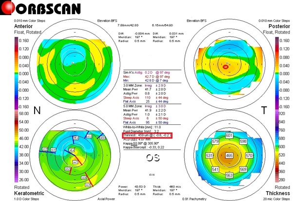
Figure 2:Postoperative Orbscan map. Corneal apex: located closely in the center of the cornea (red circle).
FIGURE 3
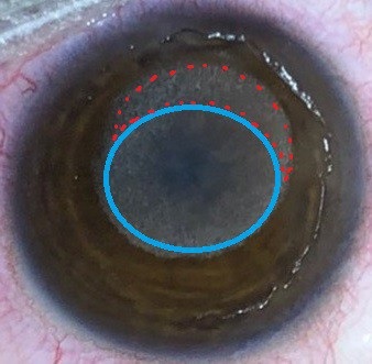
Figure 3:Laser ablation pattern. LASIK (Purple circled area) and LAK ablation pattern (Red circled area) on the thicker area of the cornea.
FIGURE 4
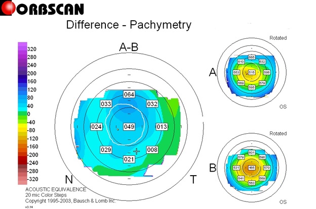
Figure 4:Orbscan Diffential pachymap between Pre- and Postoperative two months.
Figures at a glance